


Researcher(s)
- Trevor Byles, Biochemistry, University of Delaware
Faculty Mentor(s)
- Tatyana Polenova, Department of Chemistry and Biochemistry, University of Delaware
- Somayeh Zeinalilthori, Department of Chemistry and Biochemistry, University of Delaware
Abstract
Capsid interactions with host factors play critical roles in multiple steps of the HIV-1 viral lifecycle. CypA, a regulatory protein found in the cytoplasm, is a host factor packaged in the HIV-1 virions, which binds to the CA capsid protein and is required for HIV-1 infectivity.
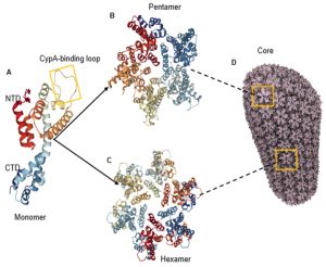
In the conical capsid, CA capsid protein is assembled from exactly 12 pentameric and a varied number of hexameric building blocks (Figure 1). The assembly into pentamers vs. hexamers is controlled by a molecular switch1. Both pentamer and hexamer forms are required for the construction of a fullerene cone shaped virion capable of nuclear import3. Recent studies have revealed that CypA binding facilitates HIV-1 ingress and viral DNA integration2. While interactions between CypA and CA hexamers are well established, whether and how CA pentamers interact with CypA is not well understood.
The Polenova lab has elucidated capsid host factor interactions using magic angle spinning (MAS) NMR spectroscopy. Whereas in vitro investigations have provided a wealth of information, performing studies within the native cellular environments is necessary. For in-cell studies of capsid host factor interactions, 19F is a powerful MAS NMR probe, given fluorine’s favorable magnetic properties, virtual absence from biological molecules (eliminating background signal), and ease of incorporation into proteins. Over the summer, we focused efforts on production of CA capsid protein for these studies. As the first step, we used transformed E. coli Rosetta cells to produce natural abundance wild type CA protein to establish protocol for fluorine incorporation with high yield. We have successfully expressed CA protein and will complete the purification as well as optimizing conditions for fluorine incorporation.
The next step of our research is the preparation of the CA-M66A variant. This variant preferentially forms pentamers1 and can maintain similar structure to that of the wild-type CA1.